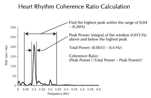
The (Power) Spectrum Average displays the amplitude of various frequency components in your heart signal. It is provided for those who enjoy frequency analysis or who are fascinated by the biological events behind heart rhythm properties.
NOTE: To view the Spectrum Average graph you will need to be running a session in emWave Pro (the Spectrum Average graph is not a feature of emWave2). It’s also available after a Session, although in a different format. Click on the arrow in the gray circle at the top of either the lower left or lower right window. Choose the Spectrum Average view.
The Spectrum Average graph represents a mathematical transformation of the heart rhythm data into its frequency components. It is provided for those who enjoy frequency analysis or who are fascinated by the biological events behind heart rhythm properties. Please Note: We won’t publish the mathematical algorithm that is used to produce the coherence ratio scores as it is proprietary IP to HeartMath only.
The amplitude on the vertical axis categorizes frequencies or cycles of repeating rhythms in the heart beat pattern. A good example of a repeating rhythm is our breathing. If we were to inhale slowly for 5 seconds and then exhale slowly for 5 seconds, this would complete one full 10-second respiratory cycle.
On a spectrum analysis graph, the frequencies (cycles) are represented by the number of times they repeat in one second. In our breathing example, one respiratory cycle was completed in 10 seconds. On a graph, this rhythm is converted to cycles per second (Hertz). A 10 second rhythm completes one-tenth (1/10) of a cycle per second, therefore the position of this event on the horizontal scale is at 0.1 (1/10 of a second), and the height, or amplitude, of the bar is determined by how deep the breath was. Faster breathing would position the spectral bar further to the right (shorter cycle), while a slower breath rate would move the spectral bar to the left (longer cycle).

In the heart rhythm data, many factors influence the recorded frequencies. The heart’s own intrinsic rhythm, breathing, emotions, as well as autonomic and other biological functions all contribute to the way the heart rate varies. This creates a complex set of frequencies all represented in the Spectrum Average. In coherence, many of these influences quiet, and the heart and breath synchronize into one frequency resulting in one dominant peak clearly defined at the coherence frequency around 0.1 Hertz.
In a state of coherence, the Spectrum Average will usually resemble a bell shaped curve with the tallest column around 0.10 Hz
.
The emWave (Power) Spectrum Average screen breaks the HRV wave into its frequency components. Each blue bar represents a sine wave at a particular frequency and amplitude. If you mathematically combined all of the sine waves represented by the blue bars you would get an approximation of the original HRV wave. Researchers have found that the blue bars in the differently colored regions indicate different states of activity in the autonomic nervous system.
There are three colored regions – the VLF (very low frequency) dark colored region on the left shows sympathetic activation. The LF (low frequency) region in the light colored middle is sometimes called the “baroreceptor region” which reflects the blood pressure control mechanisms between the heart and the brain. When in a coherent mode this indicates a synchronization of the sympathetic and the parasympathetic branches of the autonomic nervous system. The HF (high frequency) region on the right indicates parasympathetic activity. As you shift towards high coherence, the bars will focus around 0.1 Hz in this region and can get very high in amplitude. The white area on the right is not relevant to this analysis.
Most people initially will have a large peak to the left in the VLF region due to sympathetic activity. A peak around .2 Hz to the right in the parasympathetic region can appear when the user is in relaxation mode.
Note that the coherence scores do not have much of a relationship to traditional HRV parameters like SDNN Index which primarily measure HRV amplitude not how wave-like the HRV pattern is. If you are interested in generating traditional HRV measures, you’ll need to use another piece of equipment to record HRV data as the emWave program stores interpolated data; the inter-beat interval in milliseconds is stored every half second.